


Glaucoma
Q.1)What is Glaucoma ?
A condition in which there is progressive and irreversible damage to the optic nerve, (which is the nerve of sight), due to obstruction to the drainage of the fluid within the eyeball. This damage to the optic nerve causes blind spots to develop in the field of vision and if left uncontrolled, total blindness may result.
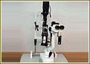
Infrastructure for the diagnosis & treatment of Glaucoma : -
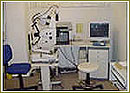
Diagnostics
 |
 |
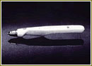 |
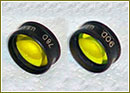
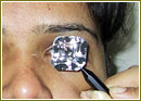
| Applanation Tonometer - to check the pressur of the eye. | 78D & 90D lens - to see the optic nerve. | Tonopen - to check the pressure of the eye |
 |
 |
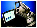 |
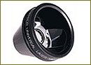
| 3 mirror GOLDMANN lens - to see the angle of the eye. | 4 mirror ZEISS gonioscopes - to see the angle of the eye | Optical Coherence Tomography (OCT III) STRATUS ZEISS: This is like a C.T. Scan of the eye. |
 |
 |
 |
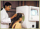
| Humphrey visual field analyzer - to measure the field of vision. | Tomey pachymeter - to measure the central corneal thickness. | TOPCON digital fundus camera - for optic disc photography. |
Medical management
Eye drops / pills have to be taken regularly.Laser treatment
The following equipments are used for laser treatment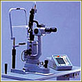
ZEISS VISULASS II -for YAG laser iridotomy.
Didode laser (iris medical) transcleral cyclophoto coagulation.
Surgical management
Includes techniques of trabeculectomy & trabeculotomy.
Modern equipments like ZEISS operating microscopes are used.
What Is The Optic Nerve?
The Optic Nerve Is A Bundle Of More Than 1 Million Nerve Fibers. It Connects The Retina To The Brain. (See Diagram Below.) The Retina Is The Light-Sensitive Tissue At The Back Of The Eye. A Healthy Optic Nerve Is Necessary For Good Vision.
Common Types Of Glaucoma
Open Angle
Open Angle (Also Called Chronic Open Angle Or Primary Open Angle) Is The Most Common Type Of Glaucoma. With This Type, Even Though The Anterior Structures Of The Eye Appear Normal, Aqueous Fluid Builds Within The Anterior Chamber, Causing The IOP To Become Elevated. Left Untreated, This May Result In Permanent Damage Of The Optic Nerve And Retina. Eye Drops Are Generally Prescribed To Lower The Eye Pressure. In Some Cases, Surgery Is Performed If The IOP Cannot Be Adequately Controlled With Medical Therapy.
Acute Angle Closure
Only About 10% Of The Population With Glaucoma Has This Type. Acute Angle Closure Occurs Because Of An Abnormality Of The Structures In The Front Of The Eye. In Most Of These Cases, The Space Between The Iris And Cornea Is More Narrow Than Normal, Leaving A Smaller Channel For The Aqueous To Pass Through. If The Flow Of Aqueous Becomes Completely Blocked, The IOP Rises Sharply, Causing A Sudden Angle Closure Attack.
While Patients With Open Angle Glaucoma Don’t Typically Have Symptoms, Those With Angle Closure Glaucoma May Experience Severe Eye Pain Accompanied By Nausea, Blurred Vision, Rainbows Around Lights, And A Red Eye. This Problem Is An Emergency And Should Be Treated By An Ophthalmologist Immediately. If Left Untreated, Severe And Permanent Loss Of Vision Will Occur In A Matter Of Days.
Secondary Glaucoma
This Type Occurs As A Result Of Another Disease Or Problem Within The Eye Such As: Inflammation, Trauma, Previous Surgery, Diabetes, Tumor, And Certain Medications. For This Type, Both The Glaucoma And The Underlying Problem Must Be Treated.
Congenital
This Is A Rare Type Of Glaucoma That Is Generally Seen In Infants. In Most Cases, Surgery Is Required.
Causes And Risk Factors
How Does Open-Angle Glaucoma Damage The Optic Nerve?
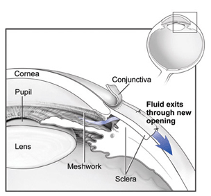
In The Front Of The Eye Is A Space Called The Anterior Chamber. A Clear Fluid Flows Continuously In And Out Of The Chamber And Nourishes Nearby Tissues. The Fluid Leaves The Chamber At The Open Angle Where The Cornea And Iris Meet. (See Diagram Below.) When The Fluid Reaches The Angle, It Flows Through A Spongy Meshwork, Like A Drain, And Leaves The Eye.
Sometimes, When The Fluid Reaches The Angle, It Passes Too Slowly Through The Meshwork Drain. As The Fluid Builds Up, The Pressure Inside The Eye Rises To A Level That May Damage The Optic Nerve. When The Optic Nerve Is Damaged From Increased Pressure, Open-Angle Glaucoma--And Vision Loss--May Result. That's Why Controlling Pressure Inside The Eye Is Important.
Does Increased Eye Pressure Mean That I Have Glaucoma?
Not Necessarily. Increased Eye Pressure Means You Are At Risk For Glaucoma, But Does Not Mean You Have The Disease. A Person Has Glaucoma Only If The Optic Nerve Is Damaged. If You Have Increased Eye Pressure But No Damage To The Optic Nerve, You Do Not Have Glaucoma. However, You Are At Risk. Follow The Advice Of Your Eye Care Professional.
How Do I Know If I Have Glaucoma?
Unfortunately Glaucoma Is Typically Associated With Painless And Progressive Loss Of Vision That May Escape Detection By The Patient. This Once Again Stresses The Importance Of A Thorough Eye History And Examination, Especially In Patients With A Family History Of Glaucoma. Only One Type Of Glaucoma Called Angle-Closure Glaucoma Is Associated With A Red, Painful Eye With Blurred Vision And Even Possibly Nausea And Vomiting. This Is Due To Very High Pressures Resulting FROM A Block In The Drainage System Of The Eye. Most Patients At Risk For This Type Of Glaucoma Have Structural Differences In Their Eye Which Could Be Identified Prior To An Attack And Preventative Treatment Could Be Performed. Rarely Do Patients With Other Types Of Glaucoma Develop Pressure High Enough To Have Pain And Redness.
Who Is At Risk For Glaucoma?
Anyone Can Develop Glaucoma. Some People Are At Higher Risk Than Others. They Include : -
- African Americans Over Age 40.
- Everyone Over Age 60, Especially Mexican Americans.
- People With A Family History Of Glaucoma.
Among African Americans, Studies Show That Glaucoma Is : -
- Five Times More Likely To Occur In African Americans Than In Caucasians.
- About Four Times More Likely To Cause Blindness In African Americans Than In Caucasians.
- Fifteen Times More Likely To Cause Blindness In African Americans Between The Ages Of 45-64 Than In Caucasians Of The Same Age Group.
A Comprehensive Dilated Eye Exam Can Reveal More Risk Factors, Such As High Eye Pressure, Thinness Of The Cornea, And Abnormal Optic Nerve Anatomy. In Some People With Certain Combinations Of These High-Risk Factors, Medicines In The Form Of Eyedrops Reduce The Risk Of Developing Glaucoma By About Half.
Medicare Covers An Annual Comprehensive Dilated Eye Exam For Some People At High Risk For Glaucoma.
Signs And Symptoms
Glaucoma Is An Insidious Disease Because It Rarely Causes Symptoms. Detection And Prevention Are Only Possible With Routine Eye Examinations. However, Certain Types, Such As Angle Closure And Congenital, Do Cause Symptoms.Angle Closure (Emergency)
- Sudden Decrease Of Vision
- Extreme Eye Pain
- Headache
- Nausea And Vomiting
- Glare And Light Sensitivity
Congenital
- Tearing
- Light Sensitivity
- Enlargement Of The Cornea
Who Diagnoses And Treats Glaucoma?
Eye Physicians And Surgeons (Ophthalmologists) Are Medical Doctors (M.D.'S), Who Have Undergone Specialized Training In ORDER To Treat Eye Diseases And To Perform Surgery. They Are Best Qualified To Diagnose Glaucoma. Some Ophthalmologists Undergo Subspecialty Training In Glaucoma And Are The Best Qualified To Treat Advanced Glaucoma. The Harvard Medical School Department Of Ophthalmology, Located At The Massachusetts Eye And Ear Infirmary, Has Many Staff Ophthalmologists Who Are Experienced In Screening Patients For Glaucoma, As Well As Glaucoma Specialists Who Are Experienced In Treating Patients Diagnosed With Glaucoma.
How Is Glaucoma Detected?
Glaucoma Is Detected Through A Comprehensive Eye Exam That Includes : -
- Visual Acuity Test. This Eye Chart Test Measures How Well You See At Various Distances. A Tonometer Measures Pressure Inside The Eye To Detect Glaucoma.
- Visual Field Test. This Test Measures Your Side (Peripheral) Vision. It Helps Your Eye Care Professional Tell If You Have Lost Side Vision, A Sign Of Glaucoma.
- Dilated Eye Exam. Drops Are Placed In Your Eyes To Widen, Or Dilate, The Pupils. Your Eye Care Professional Uses A Special Magnifying Lens To Examine Your Retina And Optic Nerve For Signs Of Damage And Other Eye Problems. After The Exam, Your Close-Up Vision May Remain Blurred For Several Hours.
- Tonometry. An Instrument (Right) Measures The Pressure Inside The Eye. Numbing Drops May Be Applied To Your Eye For This Test.
- Pachymetry. A Numbing Drop Is Applied To Your Eye. Your Eye Care Professional Uses An Ultrasonic Wave Instrument To Measure The Thickness Of Your Cornea.
Treatment
Can Glaucoma Be Treated?
Yes. Immediate Treatment For Early Stage, Open-Angle Glaucoma Can Delay Progression Of The Disease. That's Why Early Diagnosis Is Very Important.
Glaucoma Treatments Include Medicines, Laser Trabeculoplasty, Conventional Surgery, Or A Combination Of Any Of These. While These Treatments May Save Remaining Vision, They Do Not Improve Sight Already Lost From Glaucoma.
Medicines.
Medicines, In The Form Of Eyedrops Or Pills, Are The Most Common Early Treatment For Glaucoma. Some Medicines Cause The Eye To Make Less Fluid. Others Lower Pressure By Helping Fluid Drain From The Eye.
Before You Begin Glaucoma Treatment, Tell Your Eye Care Professional About Other Medicines You May Be Taking. Sometimes The Drops Can Interfere With The Way Other Medicines Work.Laser Trabeculoplasty.
Laser Trabeculoplasty Helps Fluid Drain Out Of The Eye. Your Doctor May Suggest This Step At Any Time. In Many Cases, You Need To Keep Taking Glaucoma Drugs After This Procedure.
Laser Trabeculoplasty Is Performed In Your Doctor's Office Or Eye Clinic. Before The Surgery, Numbing Drops Will Be Applied To Your Eye. As You Sit Facing The Laser Machine, Your Doctor Will Hold A Special Lens To Your Eye. A High-Intensity Beam Of Light Is Aimed At The Lens And Reflected Onto The Meshwork Inside Your Eye. You May See Flashes Of Bright Green Or Red Light. The Laser Makes Several Evenly Spaced Burns That Stretch The Drainage Holes In The Meshwork. This Allows The Fluid To Drain Better.
Like Any Surgery, Laser Surgery Can Cause Side Effects, Such As Inflammation. Your Doctor May Give You Some Drops To Take Home For Any Soreness Or Inflammation Inside The Eye. You Need To Make Several Follow-Up Visits To Have Your Eye Pressure Monitored.
If You Have Glaucoma In Both Eyes, Only One Eye Will Be Treated At A Time. Laser Treatments For Each Eye Will Be Scheduled Several Days To Several Weeks Apart.
Studies Show That Laser Surgery Is Very Good At Reducing The Pressure In Some Patients. However, Its Effects Can Wear Off Over Time. Your Doctor May Suggest Further Treatment.
Conventional Surgery.
Conventional Surgery Makes A New Opening For The Fluid To Leave The Eye. (See Diagram.) Your Doctor May Suggest This Treatment At Any Time. Conventional Surgery Often Is Done After Medicines And Laser Surgery Have Failed To Control Pressure.
Conventional Surgery Is Performed In An Eye Clinic Or Hospital. Before The Surgery, You Will Be Given Medicine To Help You Relax. Your Doctor Will Make Small Injections Around The Eye To Numb It. A Small Piece Of Tissue Is Removed To Create A New Channel For The Fluid To Drain From The Eye.
For Several Weeks After The Surgery, You Must Put Drops In The Eye To Fight Infection And Inflammation. These Drops Will Be Different From Those You May Have Been Using Before Surgery.
As With Laser Surgery, Conventional Surgery Is Performed On One Eye At A Time. Usually The Operations Are Four To Six Weeks Apart. Conventional Surgery Is About 60 To 80 Percent Effective At Lowering Eye Pressure. If The New Drainage Opening Narrows, A Second Operation May Be Needed. Conventional Surgery Works Best If You Have Not Had Previous Eye Surgery, Such As A Cataract Operation.
In Some Instances, Your Vision May Not Be As Good As It Was Before Conventional Surgery. Conventional Surgery Can Cause Side Effects, Including Cataract, Problems With The Cornea, And Inflammation Or Infection Inside The Eye. The Buildup Of Fluid In The Back Of The Eye May Cause Some Patients To See Shadows In Their Vision. If You Have Any Of These Problems, Tell Your Doctor So A Treatment Plan Can Be Developed.
Conventional Surgery Makes A New Opening For The Fluid To Leave The Eye.
How Should I Use My Glaucoma Eyedrops?
If Eyedrops Have Been Prescribed For Treating Your Glaucoma, You Need To Use Them Properly And As Instructed By Your Eye Care Professional. Proper Use Of Your Glaucoma Medication Can Improve The Medicine's Effectiveness And Reduce Your Risk Of Side Effects.
To Properly Apply Your Eyedrops, Follow These Steps : -
- First, Wash Your Hands.
- Hold The Bottle Upside Down.
- Tilt Your Head Back.
- Hold The Bottle In One Hand And Place It As Close As Possible To The Eye.
- With The Other Hand, Pull Down Your Lower Eyelid. This Forms A Pocket.
- Place The Prescribed Number Of Drops Into The Lower Eyelid Pocket. If You Are Using More Than One Eyedrop, Be Sure To Wait At Least Five Minutes Before Applying The Second Eyedrop.
- Close Your Eye OR Press The Lower Lid Lightly With Your Finger For At Least One Minute. Either Of These Steps Keeps The Drops In The Eye And Helps Prevent The Drops From Draining Into The Tear Duct, Which Can Increase Your Risk Of Side Effects
Other Related Links (Give Links To Following)
 Endocyclophotocoagulation (ECP)IridoplastyLaser Peripheral IridotomyLaser IridotmyTrabeculectomy
For more information, medical assessment and medical quote
as email attachment to
Email : - info@wecareindia.com
Contact Center Tel. (+91) 9029304141 (10 am. To 8 pm. IST)
(Only for international patients seeking treatment in India)


